解剖
空気は、外鼻孔、鼻腔、内鼻孔を通り、頬腔から声門に入ります〔Wallach 1988〕。声門は口腔内の前側に位置し、大きな獲物を飲み込むときに空気が移動できるようにします。気管は不完全な軟骨輪で構成され、心臓の高さで短い気管支に分岐します。一部のヘビの種は、血管が発達した呼吸組織からなる気管肺を持っています〔Wallach 1988〕。肺は細長い袋状の構造で、ボイドヘビを除くほとんどの種では左肺が退化しています。種とヘビが適応した環境によって、呼吸上皮で裏打ちされた右肺は、非呼吸上皮で裏打ちされた尾側の無血管の袋状の肺とつながります。水生ヘビの肺は陸生種よりも呼吸上皮が多く、長時間の無呼吸時に効率よくガス交換を行えるようになっています。
発生
飼育下のヘビでは呼吸器疾患が頻繁に診断され、罹患率と死亡率の主な原因となっています。環境条件は非常に特殊であり、飼育下ではそれを満たすことがしばしば困難で、温度や湿度が高すぎる、または低すぎる、不適切な餌、慢性的なストレスといった不適切な環境条件は、動物の免疫不全につながります。ウイルス、細菌、真菌、寄生虫など、様々な感染性病原体が検出され、呼吸器疾患と関連付けられています。
症状
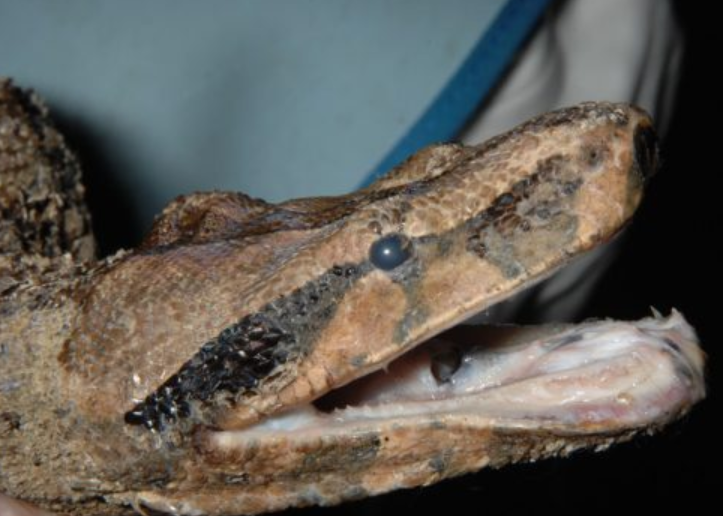
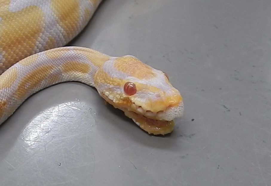
漿液性から膿性の鼻汁、喘鳴、口腔内への呼吸器分泌物の蓄積(涎)、口内炎などが一般的な症状です。重症の場合は、呼吸努力の増加や呼吸困難も現れます。ヘビは横隔膜が機能していないため、気管から呼吸器分泌物や異物を排出する機構が欠如しており、気管内に液体が溜まると気管腔が狭くなります。口腔内の粘膜あるいは声門の充血または炎症(口内炎)もみられ、食欲不振、痩削ならびに体重減少が起こります。
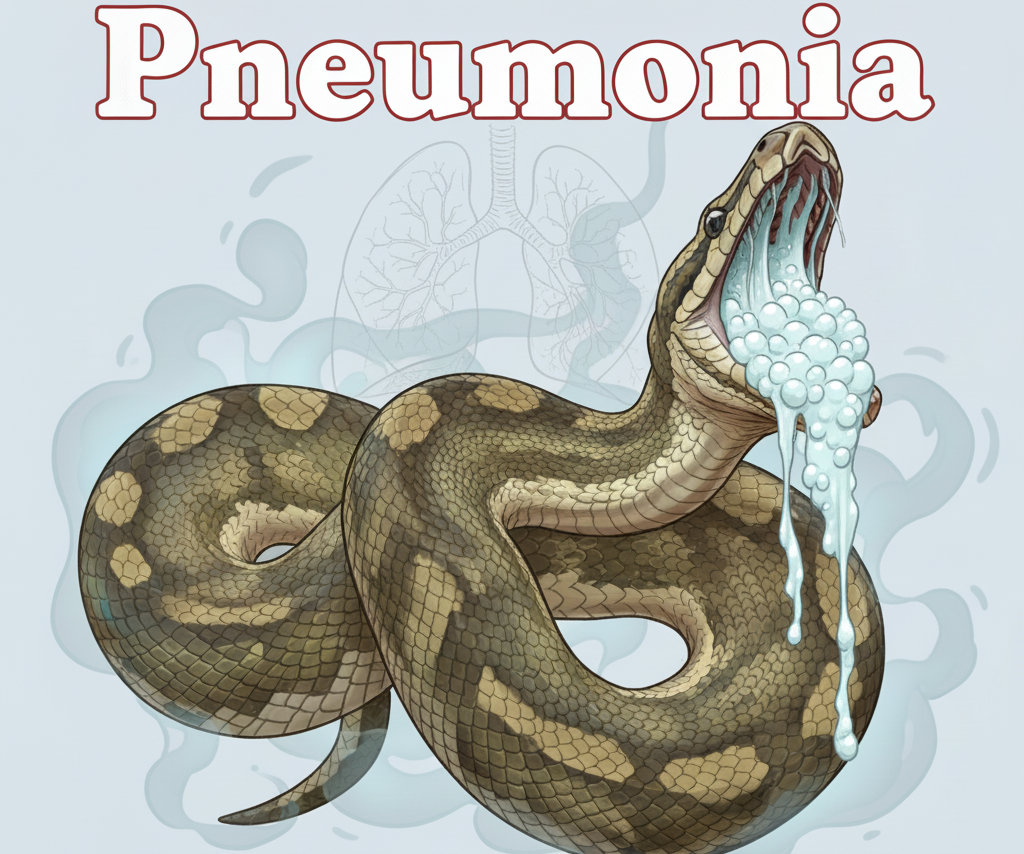
開口呼吸
ヘビの呼吸器症状は特徴的です。健康なヘビは口を閉じて呼吸をしていますが、肺炎になると開口呼吸が見られます。多くの飼い主はこの症状が見られてから、異常と気づくことが多いようです。
呼吸器分泌物の蓄積(涎)
透明、泡沫状、粘液状または粘液膿性物質の口腔からの分泌物が見られ、続いて呼吸促拍や開口呼吸などの呼吸困難症状、異常呼吸音および喘鳴(いびきのような音、「ヒューヒュー」や「ブシュー」といった異常音)も生じます。口からの分泌は鼻腔からも分泌が見られることもあります。
行動異常
樹上性のヘビでは樹木にとまるのが難しくなることもあり、呼吸困難なヘビは頭と鼻先を上に30~45度くらいに傾けた状態になり、スターゲイジングと呼ばれています。体調不良の結果、脱皮不全やアイキャップ遺残なども起こり、場合によっては突然死することもあります〔Bodewes et al.2014,Stenglein et al.2014,Uccellini et al.2014,Dervas et al.2017〕。
好発種
ヘビの肺炎は一般的で、珍しい病気ではありませんが、特にボールパイソンやボアコンストリクターに好発します。
原因
爬虫類の呼吸器疾患には、感染性および非感染性の両方の病原体が関連していることが知られています。細菌、特にグラム陰性細菌は、急性または慢性の呼吸器疾患を患う爬虫類から一般的に分離され、健康な動物にも存在する日和見細菌であることが多いです。しかし、低温飼育やビタミンA欠乏症など免疫不全の爬虫類など、特定の条件下では、これらの細菌が過剰に増殖し、主要な病原体となることがあります。他にも脱皮不全による古い皮が鼻の穴を塞いでいたり、木製チップやタバコの煙などに含まれる化学性の刺激物、不潔なケージのアンモニア臭などが呼吸器粘膜に対して刺激を与えます。爬虫類の呼吸器疾患においては、ウイルス、細菌、真菌、寄生虫などの感染性病原体が検出され、関連付けられています。多くの場合、二次的な細菌感染が一次性ウイルス性肺炎の診断を複雑化させます。適切な抗菌療法を行っても臨床症状が一時的にしか改善しない患者では、真菌、ウイルス、寄生虫因を疑う必要があり、更なる検査が必要です。
細菌
細菌
飼育下の爬虫類では細菌性肺炎がしばしば診断され、呼吸器疾患を呈する爬虫類から分離される細菌のほとんどはグラム陰性で、常在菌叢や環境中に存在します。一般的に分離される細菌には、シュードモナス属、クレブシエラ属、プロテウス属、エロモナス属(エロモナス感染症)、サルモネラ属、ブドウ球菌などです。これらは健康な爬虫類の常在菌叢の一部であるが、呼吸器疾患を呈する爬虫類の気管洗浄液からこれらの細菌が分離されることは、細菌性肺炎の兆候といえます。
真菌
真菌性肺炎はカビによる感染で、特に換気が悪い(不十分な換気)飼育ケージで起こりやすいです。
ウイルス
原因は細菌とウィルスが複雑に併発して感染していることが多いです。ヘビの肺炎では、パラミキソウイルス、アデノウイルス、オルソレオウイルス、ニドウイルス、アレナウイルスなど多くのウイルスが関与します。パラミキソウイルスは、ヘビに肺炎を起こす主な原因の一つと昔から言われていますが、野生のヘビに好発する傾向にあります。アデノウイルスは、肝炎、胃腸炎、肺炎、神経症状に関連して、オルソレオウイルスは、神経症状、胃腸炎、皮膚病変、肺炎、そして突然死に関連して検出され、特異的な症状が見られると言うより、全身に蔓延して、その結果肺炎時にも分離されます。近年はニドウイルスによる肺炎が注目されており〔Eva et al.2017〕、特にボールパイソンに多く見られるボールパイソンニドウイルス(Ball python nidovirus)という特異的なウイルスも発見されました〔Blahak et al.2020a,Mark et al.2014〕。
オーストラリアのニシキヘビからは、呼吸器症状と神経症状を引き起こす新ウイルスであるサンシャインウイルス(Sunshine virus)も発見されました(当初はパラミクソウイルス科と見なされていましたが、2016年にモノネガウイルス目内の独自のサンウイルス科に分類れました)〔Marschang et al.2013〕。ボアやニシキヘビでは封入体疾患(IBD)により慢性的な肺炎が起こり得ます(原因はレトロウイルス〔Wozniak et al.2000〕あるいはアレナウイルス〔Simard J et al.2020〕とされています)。
寄生虫
ヘビでは寄生虫による肺炎を起こすことがあり、特にヘビのWC個体では線虫である肺虫(Rhabdias spp.)と鉤虫(Kalicephalus spp.)〔Matt et al.2020,Hallinger et al.2020〕、また舌虫(Raillietiella orientalis)〔Walden et al.2020〕、肺ダニ(Entonyssus spp.)〔Fain et al.2009〕などが肺に寄生するか、発育ステージの一環で肺を移動することで、肺に損傷を与える可能性があります。
その他の原因
クラミジア〔Jacobson et al.2002〕やマイコプラズマ〔Penner et al.1997,Schmidt et al.2013〕も肺炎のヘビから検出されますが、病原性などは不明です。ヘビ(爬虫類)の声門は口底に位置するため、マウスロット(細菌性口内炎)や副鼻腔炎、または眼の感染症にかかったヘビは、肺炎を併発しやすくなります。
大半は複合感染
肺炎の原因が細菌とウイルス、それに加えて寄生虫など複合していることは、爬虫類では珍しくありません〔Jacobson 2007〕。複数のウイルス感染もあり得るため、本来病原性が高くないオルソレオウイルス 〔Hoon-Hanks et al.2020〕、あるいは呼吸器に特異的でない レトロウイルス 〔Dervas et al.2017,Blahak et al.2020〕などがニドウイルスと一緒に混合感染している例などが多く報告されています。パラミキソウイルス、アデノウイルス、オルソレオウイルスの3種のウイルスが同時に検出されたコーンスネークもいました〔Abbas et al.2011〕。ウイルス単独の感染だけでなく、二次的な細菌感染や成体の免疫などが肺炎の重症度と症状の進行に寄与する可能性があります〔Hoon-Hanks et al.2019〕。
診断・検査
X線検査
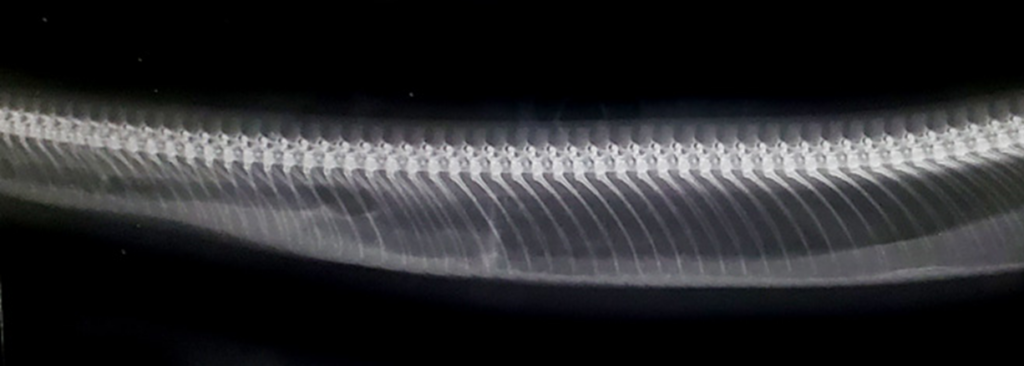
X線検査は呼吸器疾患の検出に非常に有用な診断ツールですが〔Schumacher et al.2001〕、ヘビの呼吸器系を評価する場合、細菌性肉芽腫や腫瘍などの重篤な肺の変化がない限り、X線検査では病変が検出できません。ヘビでは、細菌性肉芽腫などの肺の密度が増加した領域が、ラテラル像でよく視覚化されます。
身体検査
画像診断で評価できないことが多いため、全身状態、特に呼吸状態に注意を払う必要があります。軽度の呼吸器疾患の兆候としては、鼻汁、口腔内分泌物(涎)、眼分泌物や結膜炎などが挙げられます。安静時呼吸数の増加、呼吸困難、口を開けた呼吸、そして目に見える呼吸不全の兆候など、呼吸困難の兆候を注意して観察してください。また、開口させて声門の炎症が、肺炎の診断に有力です。これらの兆候のみで暫定的に診断することもあります。
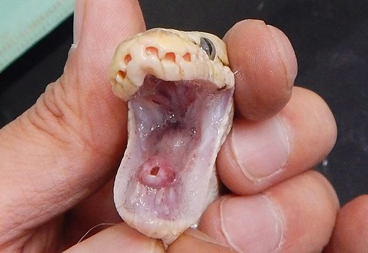
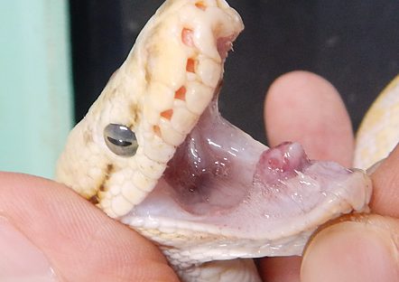
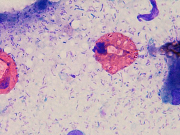
声門からの分泌物の細胞診検査で細菌や炎症細胞などが検出されます。
菌・ウイルス分離
鼻炎や上気道疾患の場合、診断用サンプルは滅菌綿棒または鼻洗浄で採取できます。採取したサンプルは適切な培養培地に移し、微生物の増殖と同定を行います。気管炎、気管支炎、肺炎などの下気道疾患が疑われる場合は、気管洗浄または肺洗浄を実施する必要があります。滅菌気管内チューブで挿管され、ヘビの大きさに応じて適切なサイズの滅菌カテーテルが気管内チューブを通して気管に挿入され、滅菌生理食塩水(3~5 mL/kg)が投与され、その後、穏やかに繰り返し吸引されます。採取した材料は、細胞学的評価、寄生虫スクリーニング、細菌および/または真菌培養、および感受性試験のために提出する必要があります。細菌は分離できますが、ウイルスは研究所でのPCR検査が行われていますが、商業検査センターでは残念ながら行われていません。
治療
治療は基本的に抗生物質を投与します。まだ食欲があり、食欲があるヘビでは餌のマウスに薬を入れて投与が可能です。食欲がない場合は注射をします。ネブライザー(噴霧治療)で、治療をすることもあります。しかし、ヘビの肺炎は多数の微生物が原因として併発しているために、単に抗生物質の投与だけで治ることもあれば、ウイルスに対する特異的な治療薬はありませんので、治らずに病状が進行するヘビもいるわけです。
【治療】エキゾチックアニマルのネブライザー治療の詳細な解説はコチラ
ポイントはコレ
・ヘビの肺炎は一般的
・ボールパイソンやボアコンストリクターに多い
・細菌や真菌、ウイルス感染による
・低温や高湿度、栄養のアンバランス、免疫力低下が発生要因
・抗生物質で治療
待望の新刊! 爬虫類の病気百科
エキゾチックアニマル臨床の第一人者 霍野晋吉が贈る、獣医師そして飼育者、ブリーダーまで、全爬虫類関係者へ送る医学バイブル

動物看護師の教科書
爬虫類好きなら持っていないといけない本!

参考文献
- Abbas MD,Marschang RE,Schmidt V,Kasper A,Papp T.A unique novel reptilian paramyxovirus,four atadenovirus types and a reovirus identified in a concurrent infection of a corn snake (Pantherophis guttatus) collection in Germany.Vet.Microbiol150:70–79.2011
- Blahak S,Jenckel M,Höper D,Beer M,Hoffmann B,Schlottau K.Investigations into the presence of nidoviruses in pythons.Virology Journal17(6).2020
- Bodewes R,Lempp C,Schürch AC,Habierski A,Hahn K,Lamers M et al.Novel divergent nidovirus in a python with pneumonia.J Gen Virol95 (Pt.11):2480-2485.2014
- Blahak S,Jenckel M,Höper D,Beer M,Hoffmann B,Schlottau K.Investigations into the presence of nidoviruses in pythons.Virology Journal17(6).2020a
- Dervas E,Hepojoki J,Laimbacher A,Romero-Palomo F,Jelinek C,Keller S et al.Nidovirus-associated proliferative pneumonia in the green tree python (morelia viridis).J Virol91:e00718‐717.2017
- Eva Dervas,a Jussi Hepojoki,a,b Andrea Laimbacher,c Fernando Romero-Palomo,a Christine Jelinek, aSaskia Keller,Teemu Smura,b Satu Hepojoki,Anja Kipar,Udo Hettzzel.Nidovirus-Associated Proliferative Pneumonia in the Green Tree Python (Morelia viridis).J Virol1.91(21).e00718-17.2017
- Fain A et al.Entonyssus squamatus spec.nov.(Acari, Entonyssidae) from the lung of the snake, Elaphe schrencki Stejneger,1925.p77-80 | Published online:17 Mar 2009
- Franke J et al.Identification and molecular characterization of 18 paramyxoviruses isolated from snakes.Virus Res28.80(1-2):67-74.2021
- Hallinger et al.Occurrence of Kalicephalus,Strongyloides,and Rhabdias nematodes as most common gastrointestinal parasites in captive snakes of German households and zoological gardens.Parasitol Res119(3):947-956.2020
- Hoon-Hanks LL,Stöhr AC,Anderson AJ,Evans DE,Nevarez JG,Díaz RE et al.Serpentovirus (nidovirus) and orthoreovirus coinfection in captive veiled chameleons(Chamaeleo calyptratus)with respiratory disease.Viruses12:1329.2020
- Jacobson E.Update on ophidian paramyxovirus infections.Proc Am Assoc Zoo Vet223.1995
- Kurath G,Batts WN et al.Complete genome sequence of Fer-de-Lance virus reveals a novel gene in reptilian paramyxoviruses.J Virol78(4).2045-56.2004
- Jacobson E,Origgi F,Heard D,Detrisac C.Immunohistochemical staining of chlamydial antigen in emerald tree boas(Corallus caninus).J Vet Diagn Invest14(6):487–494.2002
- Mark D.Stenglein,a Elliott R.Jacobson,b Edward J.Wozniak,c James F.X.Wellehan,d Anne Kincaid,e Marcus Gordon,f Brian F.Porter,et al.Ball Python Nidovirus:a Candidate Etiologic Agent for Severe Respiratory Disease in Python regius.mBio 5(5)e01484-14.2014
- Marschang RE et al.First detection of sunshine virus in pythons(Python regius) in Europe.In Association of Reptile and Amphibian Veterinarians (ARAV).Indianapolis.USA.2013
- Matt CL et al.Kalicephalus hookworm infection in four corn snakes (Pantherophis guttatus).Journal of Exotic Pet Medicine34.62-66.2020
- Penner JD et al.A novel Mycoplasma sp.associated with proliferative tracheitis and pneumonia in a Burmese python (Python molurus bivittatus).J Comp Pathol117(3):283-288.1997
- Richter GA.Characterization of paramyxoviruses isolated from three snakes. Virus Research43(1).77-83.1996
- Simard J et al.Prevalence of inclusion body disease and associated comorbidity in captive collections of boid and pythonid snakes in Belgium. PLoS One2.15(3):e0229667.2020
- Schmidt V et al.Detection of pathogens in Boidae and Pythonidae with and without respiratory disease.Vet Rec172(9):236.2013
- Schumacher J,Toal RL.Advanced radiography and ultrasonography in reptiles.Semin Avian Exotic Pet Med10:162-168.2001
- Stenglein MD,Jacobson ER,Wozniak EJ,Wellehan JFX,Kincaid A,Gordon M et al.Ball python nidovirus: a candidate etiologic agent for severe respiratory disease in python regius.MBio5:e01484-414.2014
- Uccellini L,Ossiboff RJ,de Matos REC,Morrisey JK,Petrosov A,Navarrete-Macias I et al.Identification of a novel nidovirus in an outbreak of fatal respiratory disease in ball pythons (python regius).Virol J11:144.2014
- Wozniak E,McBride J,DeNardo D,Tarara R,Wong V,Osburn B.Isolation and characterization of an antigenically distinct 68-kd protein from non-viral intracytoplasmic inclusions in Boa constrictors chronically infected with the inclusion body disease virus(IBDV:Retroviridae).Vet Pathol37(5):449-459.2020
- Walden HD et al.Case Report:Invasive Pentastomes,Raillietiella orientalis(Sambon, 1922),in a Free-Ranging Banded Water Snake(Nerodia fasciata) in North Central Florida,USA.Front Vet Sci4.7:467.2020
- Wozniak E,McBride J,DeNardo D,Tarara R,Wong V,Osburn B.Isolation and characterization of an antigenically distinct 68-kd protein from non-viral intracytoplasmic inclusions in Boa constrictors chronically infected with the inclusion body disease virus (IBDV:Retroviridae).Vet Pathol37(5):449-459.2000
- Wallach V.The lungs of snakes.Gans C,Gaunt AS.eds.Biology of the reptilia19:morphology G visceral organs.Society for the Study of Amphibians and Reptiles,St.Louis:93-295.1988